Guided DolorClast® Therapy: Successful treatment solution for supination trauma
GDT for supination trauma
Guided DolorClast® Therapy (GDT) is a new treatment concept based on combination therapies, dedicated to practitioners who want to successfully, quickly, and safely treat 90% of their patients suffering from a musculoskeletal disorder.
Supination traumas and injuries of soft tissues are very common musculoskeletal conditions. Click here and learn more about clinically proven indications and the application of GDT in musculoskeletal disorders.
Guided DolorClast®️ Therapy - 6 steps, 1 goal: pain-free patients
Step 1: Assess & Engage
Thorough taking of medical history, followed by a full passive and active functional examination of the patient is a crucial step of any successful therapy. This time, Maier consulted a retired woman who has reported a sprain trauma while doing sports. Ultrasound investigation was conducted by an orthopedist shortly after injury. The injury of the anterior fibulotalar ligament was confirmed, with significant swelling of the surrounding soft tissues and a sizable hematoma. Bone marrow edema was spotted in the medial area of the malleoli. Maier’s initial assessment two days after the injury revealed restricted gait pattern, as well as painful restriction of movement. Pain assessment scored 7 out of 10 at the VAS pain scale.
The practitioner decided on the use of the combination treatment of the DolorClast® High Power Laser and the DolorClast® Radial Shock Waves, making sure the patient was fully informed about the course of proposed therapy. Maier carefully discussed with the patient all the doubts concerning shock waves and laser therapy.
Step 2: DolorClast®️ High Power Laser
Tomas Maier has been using a high power laser device for many years in his everyday practice. That is why he decided to apply the laser treatment for the local pain relief and as further preparation for the radial shock wave therapy. Laser pretreatment allowed to deliver significantly more energy into injured tissue, which led to better clinical outcome. During each session, a 3-minutes DolorClast® High Power Laser protocol (λ=905 nm, 300 W peak power, pulsed) was administered to the painful area. Maier scheduled a 60 min break between laser and rSWT session in order to increase the amount of clinically relevant energy delivered without causing pain in the treated area.
Download our e-book and discover the potential of DolorClast® High Power Laser!
Step 3: DolorClast®️ Radial Shock Waves
Guided by the vast experience in shock waves therapy, Tomas Maier decided that the application of radial shock waves will be the most beneficial for the patient. At the beginning of treatment, parameters were set as follows: radial shock waves treatment with the 36 mm applicator, frequency 20 Hz, 8500 impulses, pressure up to max. 2.8 bar, application to the lateral and medial malleolus region reaching approximately to the lower half of the calf and the internal ankle area with the aim of pain relief and edema resorption.
Only after the first session, the level of pain decreased to VAS 1/10, the soft tissue swelling was reduced, and the gait pattern improved significantly. A compression band was applied. In addition to the combination therapy, the manual treatment called "Mobilization with Movement" ("Mobilization with Movement", MWM, Brian Mulligan) was used.
Above treatment (High Power Laser, rSWT, MWM) was repeated after two days. The patient presented no abnormalities in the gait pattern, no soft tissue swelling and assessed her pain on VAS for 1/10.
Download our e-book and discover the potential of DolorClast® Radial Shock Waves!
Finally, 2 further treatments were carried out at intervals of five days: laser pretreatment as administered earlier and RSWT with the 36 mm applicator, frequency 20 Hz, 6000 impulses on the lateral and medial malleolus area, up to 3.8 bar. The aim was a complete healing and resorption of the bone marrow edema. The patient had no difficulties with daily activities nor felt any pain.
Step 4: DolorClast®️ Focused Shock Waves
In this particular case, after the assessment of the patient’s condition, the practitioner decided to use DolorClast®️ Radial Shock Waves instead of Focused Shock Waves.
Step 5: Rehabilitation
An essential step of successful therapy is the introduction of an individually composed training program. At the beginning, the manual treatment called "Mobilization with Movement" ("Mobilization with Movement", MWM, Brian Mulligan) was introduced. It consisted of carrying out a passive mobilization by a practitioner, which is combined with an active movement of the patient. Neither of these should be painful for the patient. As the treatment progressed, the MWM technique was carried out in a functional starting position - standing while holding weights. In the end, distal fibula was taped posterior-cranially. The Mulligan tape system reduces the frequency of twisting an ankle and ensures increased gait security for the patient.
Six days after the trauma, the treatment simple coordination exercises with a low, linear load were added.
Step 6: Follow-up
The patient was reassessed at the start of each session; significant improvement (pain, mobility, range of motion) was noticed already from the second session. Eight days after the injury, a first Nordic walking round (5km) was already possible without pain and post-reaction. The patient continued to carry out the rehabilitation program to improve strength and coordination independently, as a part of her individual training. Thanks to the rapid improvement in pain and mobility, the patient's compliance was very good.
Take-home message
Get your patients going using the combination of superior technologies in rehabilitation: DolorClast® High Power Laser and DolorClast® Radial Shock Waves. Engage your patients in the 6 steps GDT protocol and manage musculoskeletal pathologies better!
Related products

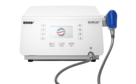
DolorClast® High Power Laser